


Wszystkie oferowane przez nasze oddziały zabiegi są wykonywane w ramach kontraktu z Narodowym Funduszem Zdrowia
![]()


Po pierwsze odpowiednia dieta
Prawidłowe odżywianie odgrywa ogromną rolę w utrzymaniu dobrego zdrowia, a tym samym wpływa na długość życia. W praktyce polega na regularnym dostarczaniu organizmowi wszystkich niezbędnych składników odżywczych w odpowiednich ilościach i proporcjach. Jeżeli zatem chcesz, aby dieta była Twoim sprzymierzeńcem, zatroszcz się o to, by twój nowy, zdrowy jadłospis zawierał: produkty zbożowe, warzywa i owoce, mleko i produkty mleczne oraz mięso, wędliny, drób, ryby i jaja. Pamiętaj, że złe nawyki żywieniowe przyczyniają się do wielu ciężkich chorób – otyłości, cukrzycy typu 2, chorób układu sercowo-naczyniowego oraz nowotworów. Z drugiej strony odpowiednie żywienie jest podstawową metodą profilaktyki i leczenia. Zatem, jedz dobrze!
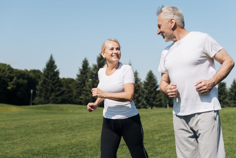
Po drugie aktywność fizyczna
Regularna aktywność fizyczna jest niezwykle istotna w życiu każdego człowieka. Umiarkowany wysiłek powoduje korzystne zmiany w układzie ruchowym. Wspomaga sprawność intelektualną. Zmniejsza napięcia nerwowe, stany depresyjne i lękowe. Poprawia jakość snu i samopoczucie. Ma pozytywny wpływ na układ hormonalny. Dobrze wpływa na układ immunologiczny, a także poprawę wydolności. Nie do przecenienia jest korzystny wpływ wysiłku fizycznego na funkcjonowanie organizmu w wieku starszym, opóźnia bowiem procesy demencji i postępowanie choroby Alzheimera. Osoby regularnie podejmujące aktywność fizyczną deklarują wyższą ocenę własnego stanu zdrowia, lepsze samopoczucie, zarówno z punktu widzenia fizycznego, jak i psychicznego oraz cieszą się zdecydowanie lepszą jakością życia. Przekonaliśmy nieprzekonanych?
Po trzecie pozytywne nastawienie
Pozytywne myślenie, pro aktywne nastawienie do świata, szacunek dla siebie i innych oraz poczucie własnej wartości to zbiór cech, które pozwalają czerpać prawdziwą radość z życia. Pozytywne myślenie jest również umiejętnością wyciągania wniosków z własnych przeżyć – również tych trudnych – ponieważ wszelkie doświadczenia mogą stać się zalążkiem korzystnej zmiany kierunku działania i mogą motywować do lepszych wyborów życiowych w przyszłości. To w jaki sposób myślimy o sobie i innych tworzy środowisko chemiczne dla komórek naszego ciała. Myśli powodują reakcję łańcuchową w organizmie. Pozytywne myślenie pomaga w tworzeniu sprzyjających harmonijnych procesów chemicznych – chociażby poprzez wydzielanie hormonów szczęścia, czyli endorfin, które aktywują układ odpornościowy. Podwyższa się także we krwi zawartość leukocytów, czyli naturalnych komórek obronnych organizmu. Nie bez przyczyny mówi się, że: „w zdrowym ciele zdrowy duch”!